Magnetic resonance imaging (MRI) is a medical procedure that uses strong magnetic field and radio waves to produce cross-sectional images of organs and internal structures of the body. MRI can give information about organs and tissues obtaining the usage of harmful X-ray radiation.
MRI is used to diagnose pathology:
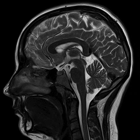
Brain and spinal cord MRI is the most informative technique used to detect the following pathological diseases:
tumors
congenital disorders
aneurism
stroke
pituitary diseases
progressive dementia
trauma of spinal cord
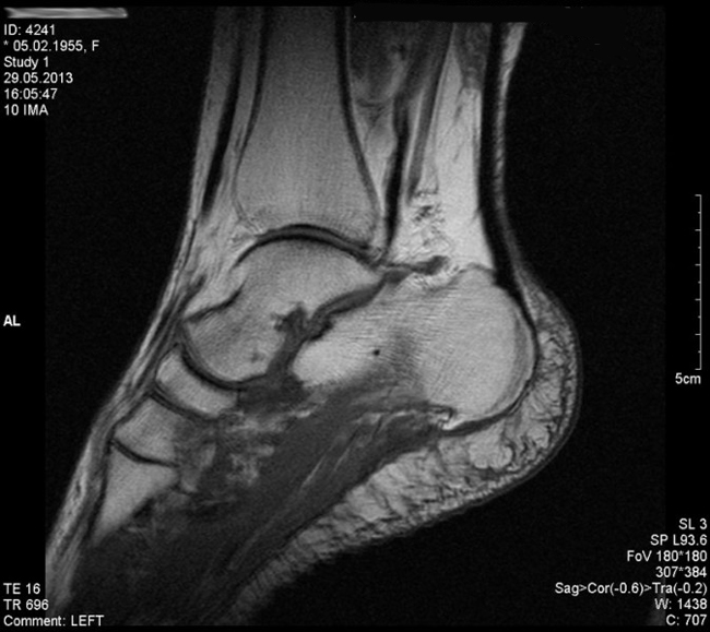
MRI of bones and joints is the basic diagnostic method to determine the following diseases:
spinal disc herniation
spondylolisthesis
spinal stenosis
arthritis
trauma of the joints
osteomyelitis
MRI of internal organs is conducted to reveal tumors and other pathologies in the following organs:
lungs
liver
kidneys
spleen
pancreas
uterus
ovaries
testicles
In addition to mammography, the MRI of mammary gland is done to diagnose cancer.
The principle of diagnosis:
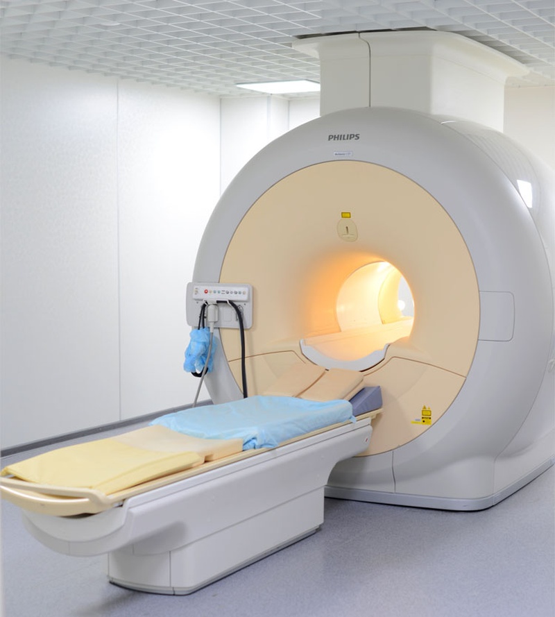
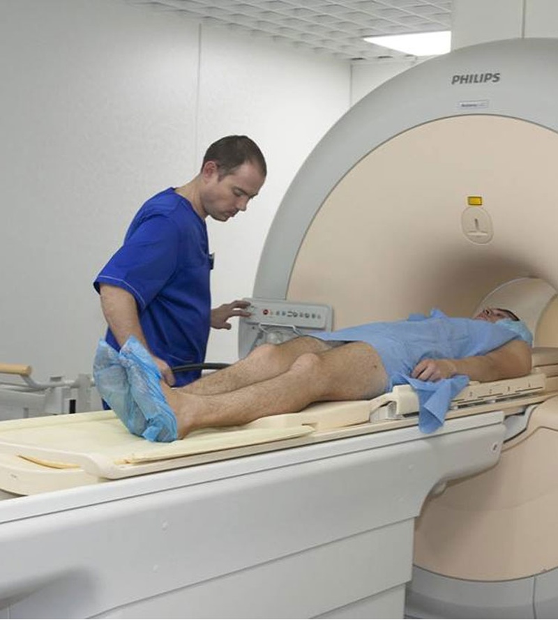
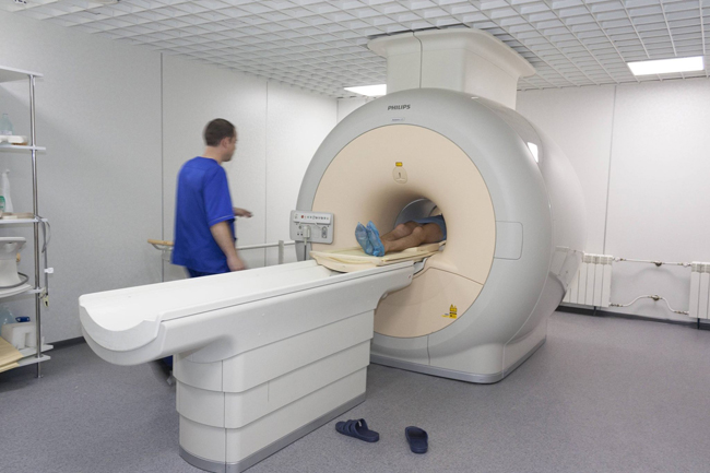
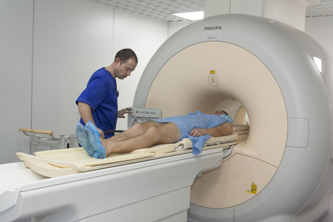
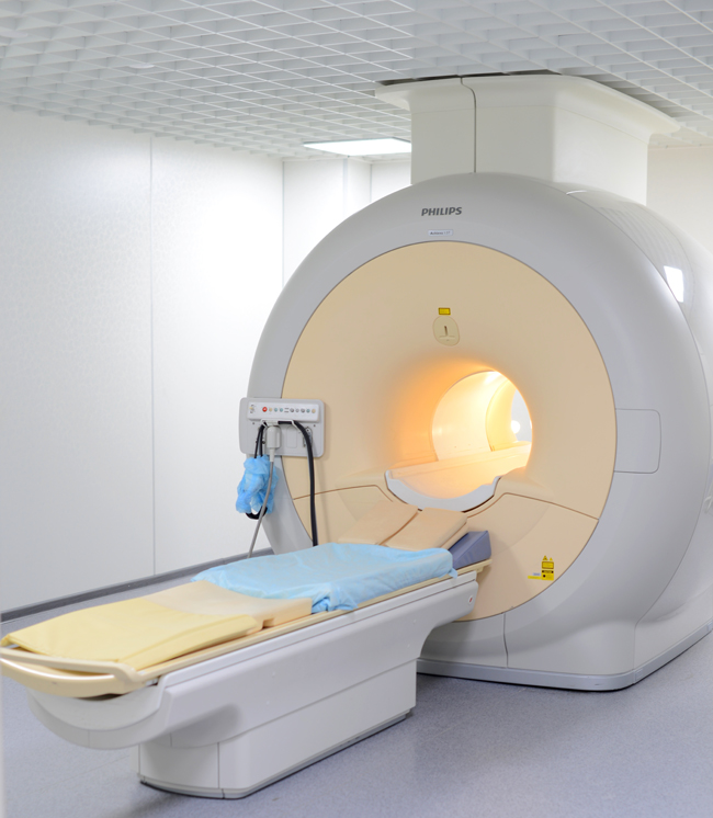
Patient is positioned on a narrow table, which is then moved inside the tunnel. While you are inside the scanner, radio waves redirect alignment of hydrogen atoms that naturally exist within the body, which is done without any chemical changes within the tissues. As the hydrogen atoms return to their usual alignment they emit energy that varies according to the type of body tissue. The MR scanner captures this energy and recreates images of the tissues based on this information. Computer processes the signals and generates a series of images, which show a small part of the body.
Benefits and risks of the MRI
Magnetic field and radio waves are absolutely harmless for the human body, as there is no damaging radiation involved.
However, the presence of a strong magnetic field means that metal objects of any kind are not permitted within the scanning room during MRI scanning.
If there are any iron-bearing objects in your body (such as vascular clips, metal fixators, metal and magnetic implants, etc.), the examination should be cancelled, as there is a high risk of their displacement, which results in damaging the surrounding tissues. Furthermore, due to the heating of the metal objects, the images produced can be distorted, leading to false interpretation.
Strong magnetic field can trouble or destroy the work of implanted electric devices, such as hearing aid or cardiac pacemaker. In order to avoid the above stated risks, it is forbidden to bring electronic items and data carriers, such as cassettes, disks, and credit cards.
Special preparation for the MRI
The patient has to give the radiologist all the necessary clinical information including previous CT and MRI reports in order to receive the required and accurate interpretation of the images. No special preparation is required before the MRI.
You can eat, drink and take medication as usual. However, if you are planning to have your abdominal cavity organs examined, you should refrain from any solid food consumption 4 hours prior to the examination. On average, the procedure is 30-45 minutes in duration, but it can sometimes last over one hour. Urinary bladder should be full when preparing for an examination of pelvic organs, however it should be empty when doing the MRI scan of other parts of the body. Makeup such as eyeshadows and mascara can cause difficulties in providing quality images for brain diagnosis. Application of make-up is therefore not recommended prior to going into the MRI procedure. In order to obtain a better visualization, any undergarments worn should be made from natural fabrics.
All jewelry and metal-containing clothing, watches, dental prosthesis, electric devices, as well as objects that contain particular elements of iron need to be removed.
Children under the age of 5 may need sedation; the process of preparation before the procedure should be discussed with anesthetist.
Make sure to inform your doctor if you:
have implanted a heart pacemaker, metallic joint prosthesis, artificial mitral value, electric devices inside body;
are pregnant;
weigh more than 120 kilograms;
have had eye surgery or intervention which have aimed to remove foreign elements;
were operated with using clips;
have metallic objects, such as, fragments after injuring, etc.
Make sure to inform your doctor if you:
• have implanted a heart pacemaker, metallic joint prosthesis, artificial mitral value, electric devices inside body;
• are pregnant;
• weigh more than 120 kilograms;
• have had eye surgery or intervention which have aimed to remove foreign elements;
• were operated with using clips;
• have metallic objects, such as, fragments after injuring, etc.
The MRI with the contrast dye
Attention! MRI with contrast dye can be offered to you in order to obtain additional information as well as help the medical staff receive an improved visibility of the images.
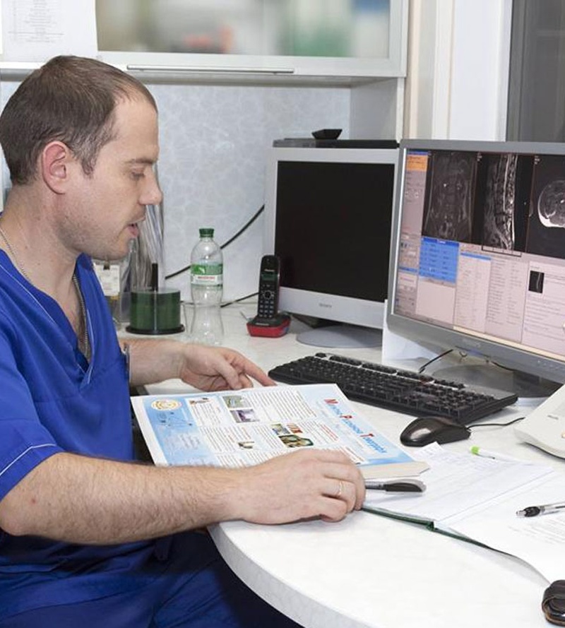
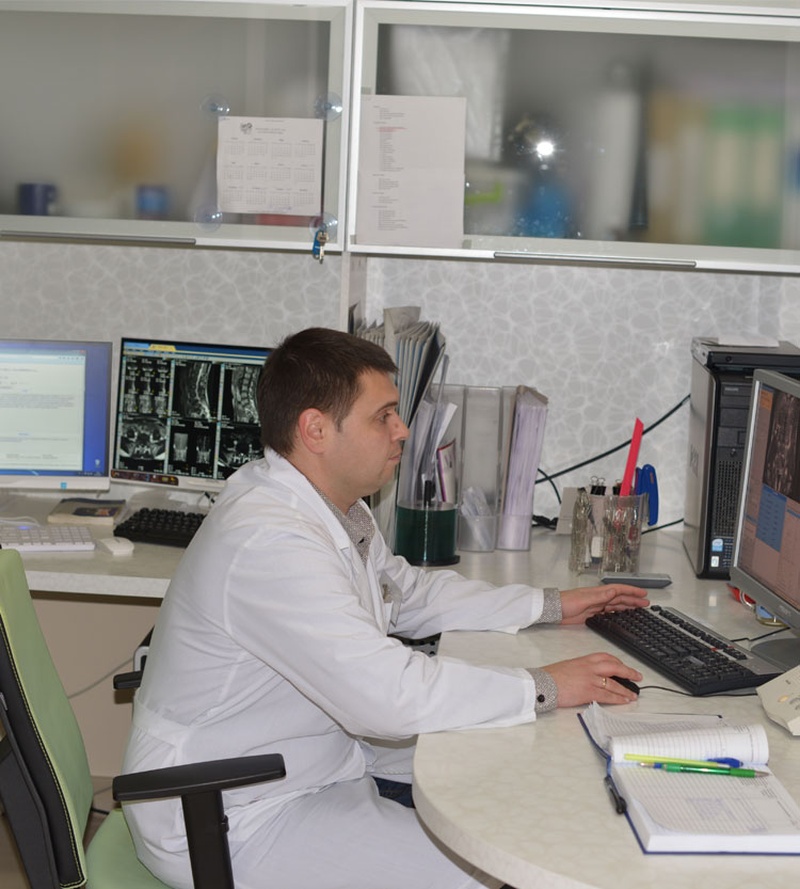
Results of the MRI
The report of the examination will be finalized after all the images are carefully studied by our professional radiologists. You will receive a CD disc with a report, film and the overall conclusion made by the doctors.

| Doctor | Name | Speciality | Work time (Tuesday) |
|---|---|---|---|
 | Мацюк Оксана Танасіївна | Рентгенологія | - |
 | Качмарський Володимир Михайлович | Рентгенологія | - |
Comments